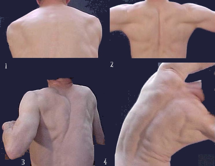
Thorax, back view
Fig. 1: Scapula in maximal lateral position. The muscles in-between the vertebral column and the medial border of the scapula present as above the "level" of the scapula. Easy to recognize are the vertebrae of the thoracic part of the vertebral column.
Fig. 2: Pressing both scapulae maximally together leads to folding of the skin and disappearance of the vertebrae of the thorax.
Fig. 3: Moving one arm forward and the other arm back induces a tissue-shift eventually over-passing the mid-line.
Fig. 4: Shows the main muscular structures of the back involved in maintaining the position of the upper body.